Search This Blog

Sunday, 5 March 2017

MAXILLARY CANINE

LABIAL ASPECT
-Crown : Narrower misodistatlly than those of maxillary central incisor
-Labial Ridge : Shallow depression mesially and distally present dividing the 3 labial lobes and middle lobe shows much greater development than other lobes
-Cervical line : Convex with convexity towards root portion
-Mesial outline : May be convex from the cervix to the centre of the mesial contact area
-Distal outline : Concave from the cervix to the distal contact area
-Cusp tip : It is on line with the center of the root
-Mesial slope of cusp is shorter than distal slope
-Root: It is narrower mesiodistally
          : Smooth and convex in all directions
          : It appears slender in form with 2 bluntly pointed apex

LINGUAL ASPECT
-Crown : Narrower lingually than labially
-Cervical line : The line may be start for short interval at this point
-Cingulum : Large and pointed all a small cusp
-Definate ridges are found below the cingulum b/w stronly developed marginal ridges
-Fossa : 2 lingual ridges and the marginal ridge
-Root : Narrower than the labial portion

MESIAL ASPECT
-Greater bulk and greater labiolingual dimension
-Crown : Wedge shaped
              : Greater dimension than distal
              :Wedge point represented by  the cusp tip
-Crest of curvature : more incisally
-Labial outline : Exhibits convexity from the crest of curvature at the cervical third, which straightens as it approaches the cusp tip
-Lingual outline : It represent convex line describing the cingulum which straightens out as the middle third is reached and it becomes covex in the incisal third
-Cervical line : Curves towards the cusp on average by approx 2.5 mm at the CEJ
-Root :Conical with tapered or blunt pointed apex
           :Curves labially towards the apical third
           :Root appears board with 2 shallow developmental depression for most of the part of root length
           :Line bisecting the cusp is labial to line bisecting the root
           :Mesial surface of the crown present convexities at all point except for a small circumscribed area above the contact area , where the surface is concave and flat b/w the area and cervical line

DISTAL ASPECT
-Cervical line : exhibits less curvature towards the cusp ridges
-Distal marginal ridges : It is heavier and more irregular in outline
-Developmental depression : Is more pronounced on the distal side of the root

INCISAL ASPECT
-Tip of the cusp : It is labial to the centre of the crown labiolingually and mesial to the center mesiodistally
-There is entire distal portion stretched to make contact with the first premolar
-The ridge of the middle labial lobe is very noticeable labially from incisal aspect
-Greater convexity at the cervical third of the crown and becoming broader and flatter at the middle and incisal third
-Outline of the cingulum : A shorter arc
-A line bisectiong the cusp and cusp ridge drawn in the mesiodistal direction is almost always straight and bisects the short arc representing the mesial and distal contact areas

PHYSIOLOGY BLOOD UNIT 11 & 12

Blood Unit 11 & 12
Platelets, Coagulation &
Bleeding Disorders

Platelet

• Platelets are produced in blood cell formation (thrombopoiesis) in bone marrow
• megakaryoblast > pro-megakaryocyte > immature megakaryocyte > megakaryocyte > Platelet
• Platelets or thrombocytes are small in size
• irregularly shaped
• non-nucleated
• 2-3 µm in diameter.
• lifespan of circulating platelets is 8 to 12 days.
• Normal Value - 1.5 to 4 lakhs/uL
• platelet production is regulated by thrombopoietin (hormone which produced by the liver and kidneys)
• Old platelets are destroyed by phagocytosis in the spleen and by Kupffer cells in the liver


• The platelet structure has 3 zones:
  - Peripheral
  - Structural
  - Organelle
• Structural zone
  - Consists of the cytoskeleton
  - The cytoskeleton forms the support for the maintenance of the platelet's discoid shape
  - Regulate contractile system that allows, upon activation, shape change, pseudopod extension, internal contraction, and release of granular constituents.


• Organelle zone
   - consists of the granules and cellular components
   - These organelles serve in the  metabolic processes of the platelet and store enzymes.
   - dense granules contain non-metabolic adenosine triphosphate (ATP) and adenosine diphosphate (ADP), serotonin, and calcium
   - alpha granules contain adhesive proteins such as fibrinogen, fibronectin, von Willebrand factor (VWF), thrombospondin.
   - alpha granules also contain growth-promoting substances such as platelet-derived growth factor (PDGF), platelet factor 4, and transforming growth factor.
   - Coagulation factors including factor V, high molecular weight, factor XI, and plasminogen activator inhibitor-1 are also present in the alpha granule.

• Membrane / peripheral zone
  - Consist of typical phospholipid bilayer membrane
  - Embedded in this structure are different kind of glycoprotein.


General function of platelet

• The function of platelets is the maintenance of hemostasis.
• Platelets helps in blood clotting.
• Wound repair
• Platelets secrete platelet-derived growth factor (PDGF).
• Granule secretion.
• Adhesion and aggregation.
• Pro-coagulation.
• Cytokine signalling.
• Phagocytosis.
• Transport of enzyme and proteins critical to clotting.
• Formation of a platelet plug to slow blood loss.
• Contraction of a clot after it has formed, which then reduces the size of the vessel break.


Platelet Adhesion

• Injured vessel get attached with vWF on the endothelium cells which attracts platelets to get attached to vWF

Platelet Activation

• Adhered platelets gets activated and changes there shape into pseudopodia, discharges granules to get more platelets attracted.

Platelet Aggregation

• Activation of phospholipase C into Phospholipase A2.
• It causes more platelet aggregation and formation of temporary haemostatic plug.
• This loose plug later gets into secondary Haemostatic Plug by fibrin.
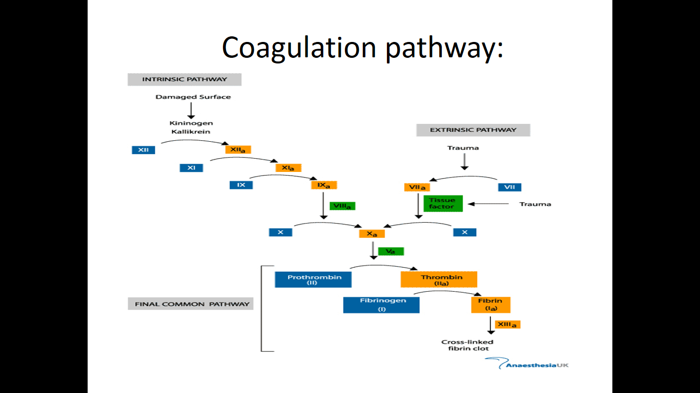
• Fibrin is activate via activation of different clotting factors.


Variations in Count

• Thrombocytosis- Increase in Platelet Count
• Causes- Administration of Epinephrine
Trauma
Removal of Spleen
• Thrombocytopenia- Decrease in Platelet Count
• Causes- Bone Marrow Depression,
Hypersplenism, Viral Infections, Leading Purpura

Types of Bleeding disorder:

• Divided into:
     - Coagulation disorder
     - Platelets disorder
• Coagulation disorder include:
     - Hemophilia
     - Von Willebrand disease • Platelet disorder include:
     - Deficiency Of Vitamin K.
     - Thrombotic Thrombocytopenic Purpura (TTP).
     - Idiopathic Thrombocytopenic Purpura (ITP).

Hemophilia

• Definition: disease associated with prolonged bleeding due to the deficiency in clotting factor.
• Hemophilia is a X-linked disease. Abnormal gene on X-chromosome
• Types of Hemophilia:
 - Hemophilia A
    -- Factor 8 deficiency, x linked disease
 - Hemophilia B
    -- Factor 9 deficiency, x-linked disease
 - Hemophilia C
    -- Factor 11 deficiency, autosomal genetic disorder
• Symptoms of hemophilia:
  - Bruising
  - Bleeds easily
  - Bleeding into a joint
  - Bleeding into the muscles
  - Bleeding from injury or bleeding in the brain
  - Other sources of bleeding (eg. Stool & urine)

Von Willebrand disease

• The most common hereditary coagulation abnormality
• Can also be acquired as a result of other medical conditions
• Due to the deficiency of von Willebrand factor (vWF)
• Von Willebrand factor  - mediates binding of glycoprotein Ib to collagen
• This binding mediate activation of platelets and formation of primary hemostasis
• Defect in this factor, resulting glycoprotein IB does not bind to collagen.
• Thus unable to activate platelets, primary hemostasis does not occur

Deficiency of Vitamin K

• Role of Vitamin K in blood coagulation:
- Important in maturation of clotting factor.
- modification of certain proteins required for blood coagulation
• If the clotting factor does not mature, it is useless in the hemostasis process.
• Factor which causes the deficiency of vitamin K - Disturbed intestinal uptake.
- By therapeutic or accidental intake of vitamin k-antagonists or very rarely.
- By nutritional vitamin k deficiency
• Some of the possible symptoms of vitamin K deficiency:
- Risk of massive uncontrolled bleeding

Defective Capillary Contractility

• This condition is known as Purpura
• Having spontaneous Haemorrhages beneath the skin, mucous membrane.

THROMBOTIC THROMBOCYTOPENIC PURPURA (TTP)

• A blood disorder that causes blood clots to form in small blood vessels around the body, and leads to a low platelet count.
• The two main types of TTP are inherited and acquired.
• In inherited TTP, the ADAMTS13 gene is faulty and doesn't prompt the body to make a normal ADAMTS13 enzyme. As a result, enzyme activity is lacking or changed.
• Acquired TTP is the more common type of the disorder. The
ADAMTS13 gene isn't faulty. Instead, the body makes antibodies (proteins) that block the activity of the ADAMTS13 enzyme.
• A lack of activity in the ADAMTS13 enzyme causes TTP.
• The ADAMTS13 gene controls the enzyme, which is involved in blood clotting.
• The enzyme breaks up a large protein called von Willebrand factor that clumps together with platelets to form blood clots.
IDIOPATHIC THROMBOCYTOPENIC PURPURA (ITP)
• Also known as immune thrombocytopenic purpura, is classified as an autoimmune disease.
• The term "idiopathic" indicates that the disease is of an unknown cause or origin: in other words, modern medicine has not yet figured out what it is.
• And the word "purpura" comes from a description of the bruisecolored skin of someone afflicted with the disease: the purple color caused by blood that leaked under the skin.

IDIOPATHIC THROMBOCYTOPENIC PURPURA (ITP)

• Idiopathic thrombocytopenic purpura is a bleeding disorder in which the immune system destroys platelets
• Persons with the disease have too few platelets in the blood
• The two types of ITP are acute (temporary or short-term) and chronic (long-lasting).
- Acute ITP generally lasts less than 6 months.
- Chronic ITP lasts 6 months or longer and mostly affects adults.
• Symptoms:
- Abnormally heavy menstruation.
- Bleeding into the skin causes a characteristic skin rash that looks like pinpoint red spots.
- Easy bruising.
- Nosebleed or bleeding in the mouth.

PHYSIOLOGY BLOOD UNIT 4

Blood Unit 4
Red Blood Cells (Erythrocytes)

Red Blood Cells

• Shape & size
- Circular Biconcave Disc
- Non-nucleated
- Diameter 7-8 um x 2.5 mm , 1 mm
- Flexible
- Number =4.5-5 x10⁶
- No nucleus, No Mitochondria, No Ribosome
- DEPEND ON Glucose Metabolism For Its Energy Source

Composition

- 62.5% water
- 35% Hb
- 2.5 %
1. Sugar
2. Lipids
3. Protein
4. Enzymes
5. Vitamins
6. Ions

Normal Values

- At birth 6-7 million/cumm
- Adults Male- 5-6 million/cumm
- Adults Female- 4.5 to
5.5 million/cumm
- Lifespan is 120 days
- 5 million/cumm is 100% RBC count clinically

Variation in Size

- Anisocytosis- Size
- Poikilocytosis- Shape
- Spherocytosis- Spherical

Genesis of RBC

• All blood cell are formed from Pluripotent hematopoietic stem cells ? committed cells:
• Committed stem cells for RBC
• Committed stem cells for WBC
• Growth of different stems cells are controlled by different growth factors

Stages of differentiation of RBC

- Stages of RBC development
   • Committed stem cell - Proerthroblast
      - basophil erythroblast
      - polychromatophil erythroblast
      - orthochromatic erythroblast
      - Reticulocytes
      - Mature erythrocytes
• Rapid RBC production  increases reticlocytes in the circulation

Proerythroblast

• No hemoglobin
• Nucleus 12 um
• Contain nucleoli

Basophil erythroblast

• Early normoblast
• Nucleoli disappear
• Show mitosis
• Cytoplasm deep blue
- Increase in RNA
• Hemoglobin starts appearing - Little Hb

Polychromatophil erythroblast

• Late normoblast
• Nucleus smaller
• Coarse Chromatin
• Hemoglobin increase
- Eosinophil Stain
• RNA - Basophil stain

Orthochromatic Erythroblast

• Normoblast
• Nucleus smaller
- Pyknosis
• Nuclear lysis and
• Nuclear extrusion

Reticulocyte

• Reticulum
• Remnant of ER & GA
- Synthesize Hb
• Few Mitochondria
• Young RBCs (34% Hb)
• 1 % of Red Cells

Transfer of RBC to Circulation

RBC pass from the bone marrow into the blood capillaries by Diapedesis (squeezing through the pores of the capillary membrane).

Erythropoiesis

- RBC development is characterize by:
   - decrease in cell size
   - disappearance of nuclus
   - appearance of haemoglobin

RBC Formation before birth

• Mesoblastic stage
    - Nucleated RBCs - Yolk sac and Mesothelial layers of the placenta -3rd week
• Hepatic stage
    - At 6 weeks - Liver form blood cells
    - Spleen + lymphoid tissues form blood cells.
• Myeloid stage
    - From the third month onwards - the bone marrow gradually becomes the principal source of the RBCs
    - Last month - Bone marrow exclusively

RBC Formation after birth

• The bone marrow - all bones - 5 years
• Marrow of the long bones (except for the proximal humerus and tibia)
    - No more red blood cells after = age 20 years.
• Most red cells continue to be produced in the marrow of the membranous bones, such as - Vertebrae, Sternum, Ribs, and Ilium.


Regulation of RBC production 

• Erythropoiesis is stimulated by erythropoietin hormone produced by the kidney in response to hypoxia (low oxygen in the blood)
• Hypoxia caused by:
   - Low RBC count (Anaemia)
   - Hemorrhage
   - High altitude
   - Prolong heart failure
   - Lung disease


Erythropoietin

• Glycoprotein
• 90% from renal cortex 10% liver
• Stimulate the growth of early stem cells
• Does not affect maturation process
• Can be measured in plasma & urine
• High levels of erythropoietin
   -anemia
   -High altitude
   -Heart failure
   -Lung Disease
(Result in polycythemia)


RBC Indices

• RBC indices include:
   - Mean Corpuscular Volume (MCV)
   - Mean Corpuscular Hemoglobin (MCH)
   - Mean Corpuscular Hemoglobin Concentration (MCHC)
   - RBC Distribution Width (RDW)
   - Colour Index (CI)

MCV

• Mean cell volume
• MCV is average size of RBC
• MCV  =   Hct x 10/RBC (millions)
• If 80-100 fL, normal range, RBCs considered normocytic
• If < 80 fL are microcytic
• If > 100 fL are macrocytic
• Not reliable when have marked anisocytosis

MCH

• MCH is average weight of hemoglobin per RBC.
• MCH = Hgb x 10/RBC (millions)

MCHC

• MCHC is average hemoglobin concentration per RBC
• MCHC = Hgb x 100 Hct (%)
• If MCHC is normal, cell described as normochromic
• If MCHC is less than normal, cell described as hypochromic
• There are no hyperchromic RBCs

RDW

• Most automated instruments now provide an RBC Distribution Width (RDW)
• An index of RBC size variation
• May be used to quantitate the amount of anisocytosis on peripheral blood smear
• Normal range is 11.5% to 14.5% for both men and women

CI

• Ratio of Hb to RBC
• Insignificant index as normal range is very wide for RBC
• CI- Hb%/RBC%
• Normal Range is 1 (0.85-1.15)

Anemia

• Anemia (An-without,emia-blood)is a decrease in the RBC count, hemoglobin and/or
Hematocrit values resulting in a lower ability for the blood to carry oxygen to body tissues .


Grading

• Mild Anemia- Hb 8-12 gm%
• Moderate Anemia- Hb 5-8 gm%
• Severe Anemia- Hb less than 5 gm%


TYPES OF ANEMIA

Based on clinical picture
* Iron deficiency anemia.
* Megaloblastic anemia.
* Pernicious anemia.
* Hemorrhagic anemia.
* Hemolytic anemia.
  -Thalassemia anemia
  -Sickle cell anemia
* Aplastic anemia
* Erythroblastosis Foetalis
* G-6PD Anemia- Decrease in Glutathione Production

• Iron deficiency anemia
   - excessive loss of iron .
   - Women are at risk. ---- For menstrual blood and growing fetus.
• Megaloblastic anemia
   - Less intake of vitamin B 12  and folic acid.
   - Red bone marrow produces abnormal RBC.
• Pernicious anemia
   - Inability of stomach to absorb vitamin B 12 in small intestine.
• Hemorrhagic anemia
  - Excessive loss of RBC through bleeding,stomach ulcers,menstruation
• Hemolytic anemia
  - RBC plasma membrane ruptures.
  - may be due to parasites,toxins,antibodies.
• Thalassemmia
  - Less synthesis of hemoglobin .Found in population of Mediterranean sea.
• Sickle cell anemia
  - Hereditary blood disorder, characterized by red blood cells that assume an abnormal, rigid, sickle shape.
• Aplastic anemia
  - destruction of red bone marrow .
  - caused by toxins,gamma radiation.
• Normochromic, normocytic anemia (normal MCHC, normal MCV).These include:
  - anemias of chronic disease
  - hemolytic anemias (those characterized by accelerated destruction of RBC's)
  - anemia of acute hemorrhage
  - aplastic anemias (those characterized by disappearance of RBC precursors from the marrow)
• Hypochromic, microcytic anemia (low MCHC, low MCV).These include:
  - iron deficiency anemia
  - thalassemias
  - anemia of chronic diseases
• Normochromic, macrocytic anemia (normal MCHC, high MCV).These include:
  - vitamin B12 deficiency
  - folate deficiency


RISK FACTORS

• Poor socio economic class
• Multiparity
• Teenage pregnancy
• Menstural problem

SIGNS OF ANAEMIA 

• Brittle nails
• Koilonychia (spoon shaped nails)
• Atrophy of the papillae of the tongue
• Angular stomatitis
• Brittle hair
• Dysphagia and Glossitis
• Plummer vinson/kelly patterson Symptoms & Signs


PHYSIOLOGY BLOOD UNIT 3

Introduction

• The main function of red blood cell
• Transfer of O2 from lungs to tissue
• Transfer of CO2 from tissue to lungs
• To accomplish this function red cells has haemoglobin (Hb)
• Each red cell has 640 million molecules of Hb

The red, oxygen carrying pigment in the RBCs is Haemoglobin. • Structure: 2 parts : heme + globin
• Globin: four chains.
• Heme: porphyrin ring with central iron. Iron is the site of attachment with O2.
• There are 4 heme groups each attached to on globin chain. So one Hb molecule can carry up to 4 O2 molecules.
• According to sequence of amino acids in the primary structure of each chain, there are four types of chains; α, β, γ and δ.

Synthesis

• Synthesis begins in proerythroblast
    - 65% at erythroblast stage
    - 35% at reticulocyte stage
• Haem & globin produced at two different sites in the cells
     - Haem in mitochondria
     - Globin in polyribosomes


Normal Values

• The hemoglobin level is expressed as the amount of hemoglobin in grams (gm) per deciliter (dL) of whole blood, a deciliter being 100 milliliters.
• The normal ranges for hemoglobin depend on the age and, beginning in adolescence, the gender of the person. The normal ranges are:
• Newborns: 17 to 22 gm/dL
• One (1) week of age: 15 to 20 gm/dL
• One (1) month of age: 11 to 15 gm/dL
• Children: 11 to 13 gm/dL
• Adult males: 14 to 18 gm/dL
• Adult women: 12 to 16 gm/dL
• Men after middle age: 12.4 to 14.9 gm/dL
• Women after middle age: 11.7 to 13.8 gm/dL

Types of Hb

• Hb A or HbA1: is the normal Hb in adults represents about 97% of total Hb. it is composed of 2 a and 2 ß chains.
• HbA2: minor adult Hb, comprised 3% of normal adult Hb. Composed of 2 a and 2 δ chains
• HbF(fetal Hb): is the main Hb during fetal life and about 60% of normal Hb at birth then disappear gradually. It is composed of 2a and 2 γ chains.
• Hb F has greater affinity for O2 than HbA so ensure O2 transfer  from maternal circulation to fetus RBCs through placenta.

Functions of Haemoglobin

• Oxygen delivery to the tissues and CO2 to lungs
• Act as excellent acid-base buffer.
• Helps in vasodilation by binding to NO.
• Reaction of Hb & oxygen
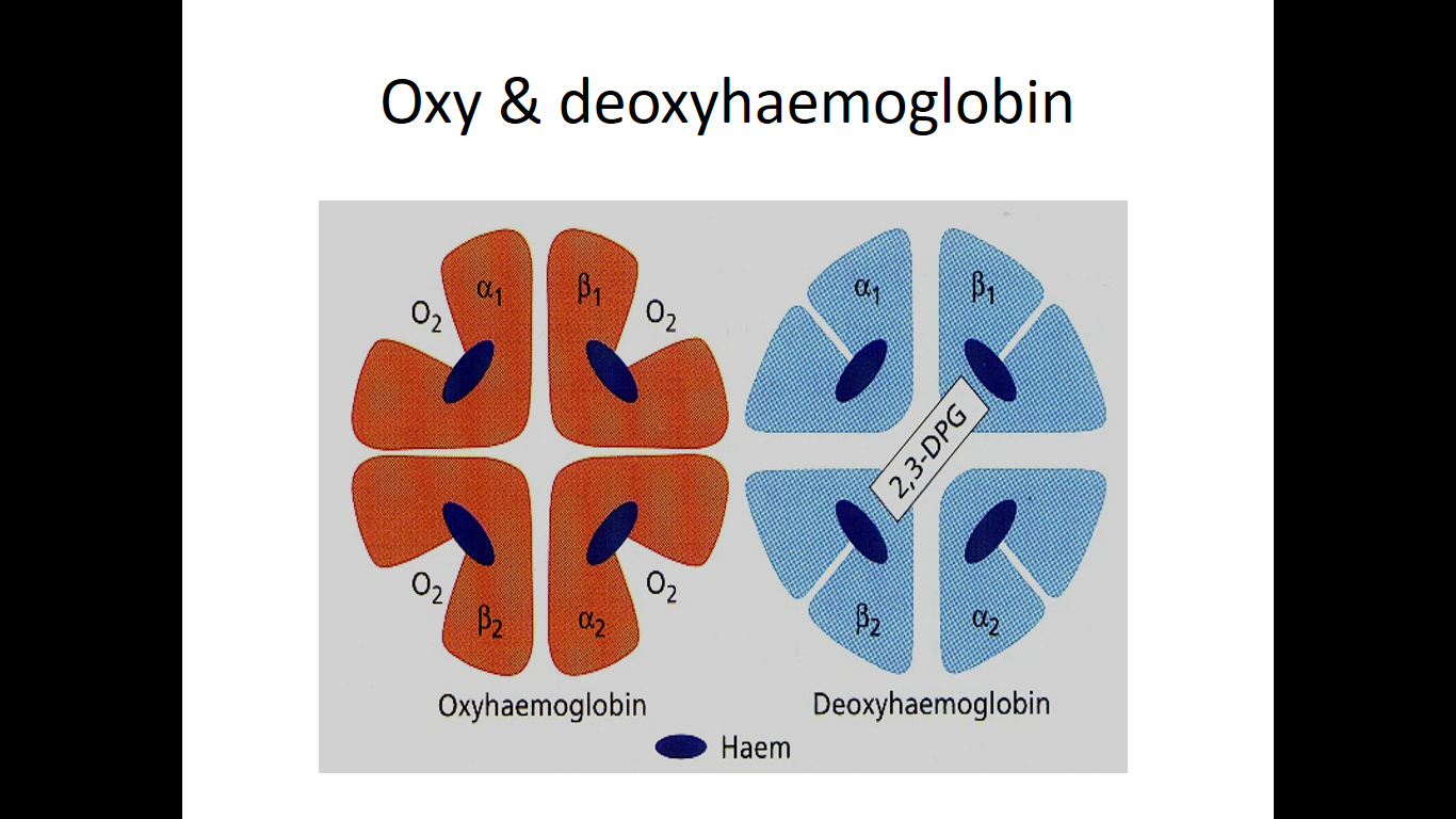
    - One Hb can bind to four O2 molecules
    - Less than .01 sec required for oxygenation
    - Oxygenation
    - b chain move closer when oxygenated
    - When oxygenated 2,3-DPG is pushed out
    - b chains are pulled apart when O2 is unloaded, permitting entry of 2,3-DPG resulting in lower affinity of O2


Oxygen-haemoglobin dissociation curve

• O2 carrying capacity of Hb at different Po2
• Sigmoid shape
     - Binding of one molecule facilitate the second molecule binding
     - P 50 (partial pressure of O2 at which Hb is half saturated with O2) 26.6mmHg


• The normal position of curve depends on
    - Concentration of 2,3-DPG
    - H+ ion concentration (pH)
    - CO2 in red blood cells
    - Structure of Hb

•  Right shift (easy oxygen delivery)
    - High 2,3-DPG
    - High H+
    - High CO2
    - HbS

• Left shift (give up oxygen less readily)
    - Low 2,3-DPG
    - HbF

Different Conjugated Hb

• Carbamino Hb- Reaction of CO2 with Hb
• Carboxy Hb or Carbon Monoxy Hb- CO reacts with Hb. Affinity of Hb for CO is 250 times  more than O2 which may affect O2 carrying capacity.
• Methaemoglobin- Exposure to various drugs or oxidising agent such ferrous to Ferric.
(Oxidation)

Mutations in hemoglobin (hemoglobinopathies:

1- Sickle cell anemia (Hb S disease):
It is a genetic disorder of blood caused by mutation in ß-globin chain resulting in the formation of Hb S. The mutation occurs in 6th position of ß-chain where glutamic acid is replaced by valine (non polar). RBCs assume sickle-shaped leading to fragility of their walls and high rate of hemolysis.


Such sickled cells frequently block flow of blood in narrow capillaries and block blood supply to tissue (tissue anoxia) causing pain and cell death.
Note: The lifetime of erythrocyte in sickle cell is less than 20 days, compared to 120 days for normal RBCs.

Patients may be :
- Heterozygotes (Hb AS): mutation occurs only in one ß-globin chain. These patients have sickle cell trait with no clinical symptoms and can have normal life span.
Or: Homozygotes (Hb SS): mutation occurs in both ß-globin chain with apparent anemia and its
symptoms

2- Hb C disease: Like HbS, Hb C is a mutant Hb in which glutamic acid in 6th position of ß-chain is replaced by lysine. RBCs will be large oblong and hexagonal.
The heterozygous form (HbAC) is asymptomatic.
The homozygous form (Hb CC) causes anemia, tissue anoxia and severe pain.

3- Thalassemia: A group of genetic diseases in which a defect occur in the rate of synthesis of one or more of Hb chains, but the chains are structurally normal. This due to defect or absence of one or more of genes responsible for synthesis of a or ß chains leading to premature death of RBCs.

Types:
ß -thalassemia: When synthesis of ß chains is decreased or absent.There are two copies of the gene responsible for synthesis of ß chains. Individuals with ß globin gene defects have either :
   
      -ß -thalassemia minor (ß -thalassemia trait) : when the synthesis of only one ß -globin gene is defective or absent. Those individuals make some ß chains and usually not need specific treatment.
      -ß -thalassemia major ( Cooley anemia): if both genes are defective. Babies will be severely anemic during the first or second year of life and so require regular blood transfusion. Bone marrow replacement is more safe treatment.
      -a-thalassemia: in which synthesis of a globin chain is defective or     absent. There are four copies of gene responsible for synthesis of a globin chains so patients may have:
-
i - Silent carrier of a-thalassemia with no symptoms: if one gene is defective
ii- a-thalassemia trait: if two genes are defective.
iii- Hb H disease: if 3 a globin genes are defective, with mild to moderate anemia. The produced Hb will be ß4 which is called HB H. Oxygen delivery to tissues will be blocked because Hb H (ß4 ) has high affinity to O2 and not deliver it to tissues.
iv- Hydrops fetalis: when all 4 a globin genes are defective. It causes fetal death because a globin chains are required for synthesis of Hb F.

HISTOLOGY SLIDES


Saturday, 4 March 2017

PHYSIOLOGY BLOOD UNIT 2

PHYSIOLOGY BLOOD UNIT 2

Introduction

Proteins present in plasma of Human Blood are mixture of simple proteins, glycoprotein, lipoproteins and other conjugated proteins together called as "Plasma Protein".
• In Embryo- they originate from mesenchymal cells with mainly Albumin as major Protein
• In Adults - Albumin & Fibrinogen from liver - Globulin from plasma cells, lymphocytes

Plasma Proteins

• Normal level of Plasma Protein concentration is 6.4-8.3 g/dL of Blood.
• It is the main contributor to osmotic pressure of the blood and it functions as a carrier molecule for molecules with low water solubility such as lipid soluble hormones, enzymes, fatty acids, metal ions, and pharmaceutical compounds.
• Degradation:
  -Hepatocytes, mononuclear  phagocytic system

Types of plasma proteins

1. Albumin - 55%
2. Globulins - 38%
 a-globulins
 b-globulins
 g-globulins
3. Fibrinogen - 7%
4. Some amount of Prothrombin

'Under different pathological conditions the protein levels depart from the normal range.
proteins
size.

Albumin

* ? 55% of the total plasma protein
* Types- Pre-Albumin & Albumin
* Pre-Albumin- Binds Thyroxine & Triiodothyronine
* Albumin
* Functions:
    • Maintenance of colloidal osmotic pressure • Protein reserve, the source of amino acids
    • Transport of:
       -steroid hormones
       -free fatty acids
       -bilirubin
       -drugs (sulfonamides, aspirin)
        -Ca2+
        -Cu2+

Causes of Albumin Deficiency

• Liver diseases (cirrhosis) - decrease in the ratio of albumin to globulins
• Protein malnutrition
• Excessive excretion by kidneys (renal disease)
• Mutation causing analbuminemia (affects splicing)

Globulin

•  ~38%% of the total plasma protein
Types -
• Glycoprotein- Carbohydrate plus protein
• Lipoprotein - lipid plus protein (Alpha-2 Globulin)
1. HDL
2. LDL
3. VLDL
4. Chylomicrons
• Transferrin
• Haptoglobins
• Ceruloplasmin
• Fetuin
• Coagulation factors
• Angiotensinogen
• Haemagglutins- Antibodies againts RBCs
• Immunoglobulin (Ig)

Low Density Lipoprotein

LDL are formed in the blood from Immediate Density .

Lipoprotein and in liver from IDL.

LDL are enriched in cholesterol and glycerides.

LDL is the major carrier of cholesterol (transport cholesterol to peripheral tissue).

High serum levels of cholesterol cause disease and death by contributing to development of  atherosclerosis.

Cholesterol which is present in the form of the LDL is so-called "bad cholesterol."

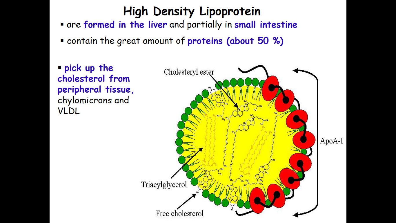
Cholesterol in the form of HDL is referred to as "good cholesterol" .

HDL functions as a shuttle that moves cholesterol throughout the body.

LDL/HDL Ratio

The ratio of cholesterol in the form of LDL to that in the form of HDL can be used to evaluate susceptibility to the development of atherosclerosis.

For a healthy person, the LDL/HDL ratio is 3.5 to 1

VLDL

• are formed in the liver
• contain 50 % of TriGlycerides and 22 % of cholesterol
• two lipoproteins - apo B-100 and apo E
• the main transport form of TGs synthesized in the organism (liver)
• deliver the TGs from liver to peripheral tissue (muscle for energy, adipose for storage)

Chylomicrons

• are the largest lipoproteins (180 to 500 nm in diameter)
• are synthesized in the ERof intestinal cells
• contain 85 % of TGs (it is the main transport form of dietary TGs).
• are present in blood only after feeding

Transferrin 

• Transferrin is a ß-globulin
• Regulates & Control iron absorption from GIT
• Controls the level of free Iron
• Protects against iron intoxication • Transferrin levels are decreased in:
  - liver disease (e.g. cirrhosis)
  - Chronic infections
  - Nephrosis
  - Congenital atransferrinaemia
• Increased serum transferrin levels occur during increased transferrin synthesis caused as a result of iron deficiency anemia

Haptoglobin (Hp) 

* a2- globulin
* Exists in 3 polymorphic forms ??Functions:
* Binds free hemoglobin and delivers it to the reticuloendothelial cells
* Prevention of loss of free Hb
* Prevents loss of Iron through Urinary Excretion

Cerruloplasmin

* Conc. in plasma: 300 mg?l
* Functions:
    - carries 90% of copper in plasma (copper - cofactor for a variety of enzymes)
    - 1 molecule binds 6 atoms of copper binds copper more tightly than albumin that carries other 10% of plasma copper -> albumin may be more important in copper transport (donates copper to tissues more readily)

Fibrinogen

• Glycoprotein, belongs to b2-globulins (Mr 340 000)
• Concentration in plasma - 1.5 - 4.5 g/l
• component of the coagulation cascade - fibrin precursor

Acute-phase proteins

• Inflammation also induces high systemic levels of acute-phase proteins. In acute inflammation, these proteins prove beneficial, however in chronic inflammation they can contribute to amyloidosis. These proteins include C-reactive protein, serum amyloid A, and serum amyloid P, vasopressin, which cause a range of systemic effects including:
• Fever
• Increased blood pressure
• Decreased sweating
• Malaise
• Loss of appetite

C-reactive protein (CRP)

• C-reactive protein (CRP) is a protein found in the blood, the levels of which rise in response to inflammation (an acute-phase protein).
• CRP is synthesized by the liver in response to factors released by fat cells (adipocytes)

Serum amyloid A (SAA)

• Serum amyloid A (SAA) proteins are a family of apolipoproteins associated with high-density lipoprotein(HDL) in plasma. These proteins are produced predominantly by the liver.
• These proteins have several roles, including the transport of cholesterol to the liver for secretion into the bile, the recruitment of immune cells to inflammatory sites, and the induction of enzymes that degrade extracellular matrix

Functions of plasma proteins

• Transport of substances : e.g.
- albumin - fatty acids, bilirubin, calcium, drugs
- transferin - iron
- cerulplasmin - copper
- lipoproteins - lipids
- haptoglobin - free hemoglobin
- thyroxin binding globulin - thyroxin

Functions of plasma proteins (cont.)

• Osmotic regulation:
- Plasma proteins are colloidal and non-diffusable and exert a colloidal osmotic pressure which helps to maintain a normal blood volume and a normal water content in the interstitial fluid and the tissues.
- Albumin content is most important in regulation of colloidal osmotic or oncotic pressure.
- Decrease in albumin level results in loss of water from blood and its entry into interstitial fluids causing edema.

• Catalytic function (enzymes):
- e.g lipases for removal of lipids from the blood

• Protective function:
- Immunoglobulins combine with foreign antigens and remove them.
- Some proteins increase during acute phase and protect the body. E.g. a1-antitrypsin, a2-macroglobulins.

• Blood clotting:
- Many factors are involved in clotting mechanism and prevent loss of excessive amount of blood; e.g. clotting fibrinogen, factors IX, VIII, thrombin, etc.
- An excess of deficiency leads to a disease; e.g. hemophilia, thrombus formation

• Anticoagulant activity (thrombolysis): - Plasmin breaks down thrombin and dissolves the clot
• Buffering capacity: - Proteins in plasma help to maintain acid-base balance
• Useful to maintain viscosity of Blood
• Maintain Systemic Arterial Blood Pressure
• Provides stability to blood- Prevent Rouleaux formation